Skin Checks

Some skin cancers are obvious and many people will identify them themselves. Others are more subtle and difficult to recognise with the naked eye. Skin cancers are more common in sun exposed areas but they can occur anywhere including parts of the body that do not see the sun. It is important to see a doctor from time to time for a full body skin check. Different people are at different risks so the frequency of these checks may vary for everyone. Your doctor will guide you on a good time frame.

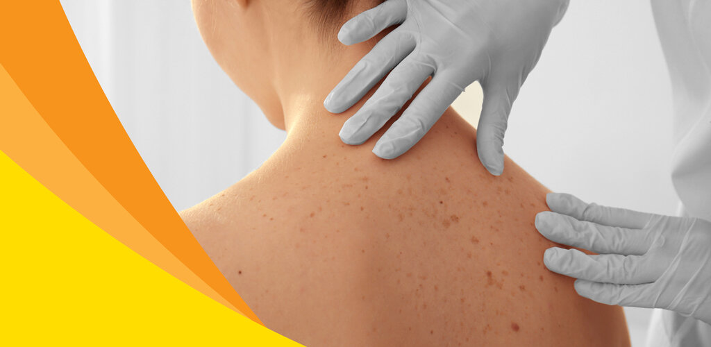
Skin checks are generally considered screening, that is the aim is to identify something you were not aware of. Sometimes if you present with a suspicious lesion the doctor may opt to do a full skin check as well. As skin cancers can occur anywhere a skin check is best performed in your underwear. You can cover with a sheet if you feel more comfortable. The doctor will examine your skin from head to toe with the aid of a dermatoscope. This is a special device that uses polarized light and magnification to see into the skin and make diagnosis easier. 

Occasionally the doctor may take photos of suspicious lesions. They can then enlarge them on a computer screen. They may decide to review the lesion in a few months or if it is more suspicious have the lesion biopsied or even excised. This generally occurs on another day.

We do ask when you come for a skin check to please refrain from wearing makeup, creams or moisturisers as this can make it more difficult to properly view your skin. But of course deodorant is fine. Occasionally skin cancers can grow beneath your finger and toenails so it is best to have any nail polish removed also. 

Whilst it can be a little daunting having your skin examined for some people we do try to make you as comfortable as possible and a positive experience.


Total Body Photography

People with a very large number of moles on their skin, particularly those that vary in size and shape are at a higher risk of melanoma. Generally melanomas are new lesions that exhibit change over time. The difficulty can be ‘picking the needle in the haystack’ as the melanoma may blend with the other moles. 

Some high risk patients may benefit from total body photography. This involves taking several photographic images of different body parts to build a representation of the full body. These can be used as reference points to detect any change for the patient when doing self checks at home or for the doctor whilst performing routine skin checks.

It is best if the photographs are updated every few years.

Services-MidPageImage_MansBack-1260x973.jpg

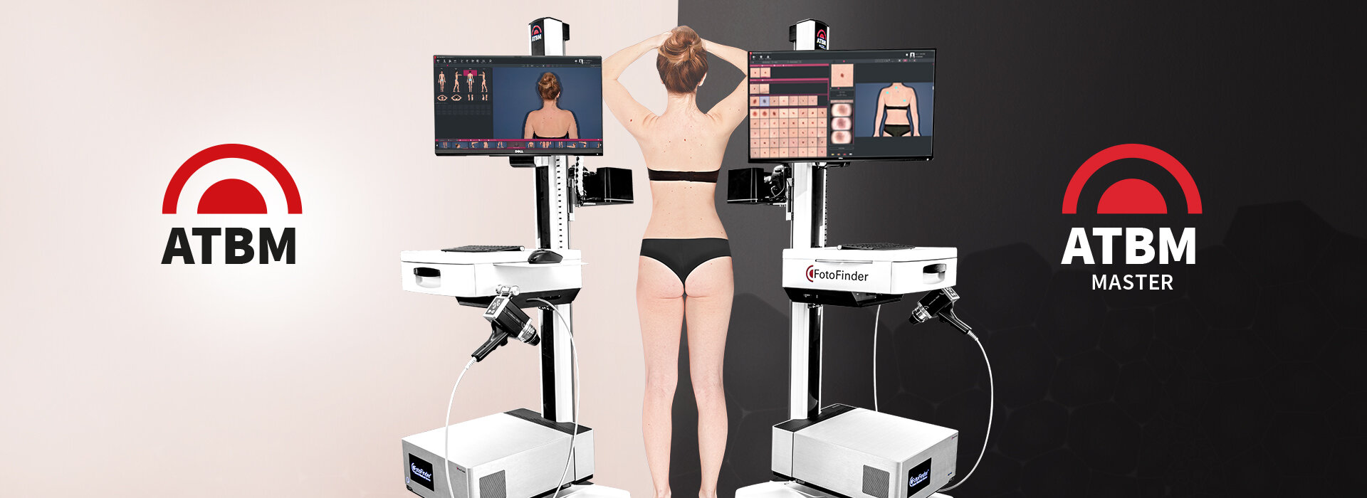
Fotofinder Automated Total Body Photography

Early detection of melanoma is the key to improving prognosis. We know that 70% of melanomas are new lesions not arising from an existing mole whilst other melanomas will exhibit change over time. Identifying these new and changing lesions in high risk patients that have a very large population of moles can be difficult. Total body photography and body mapping has been used to help detect these changes. 

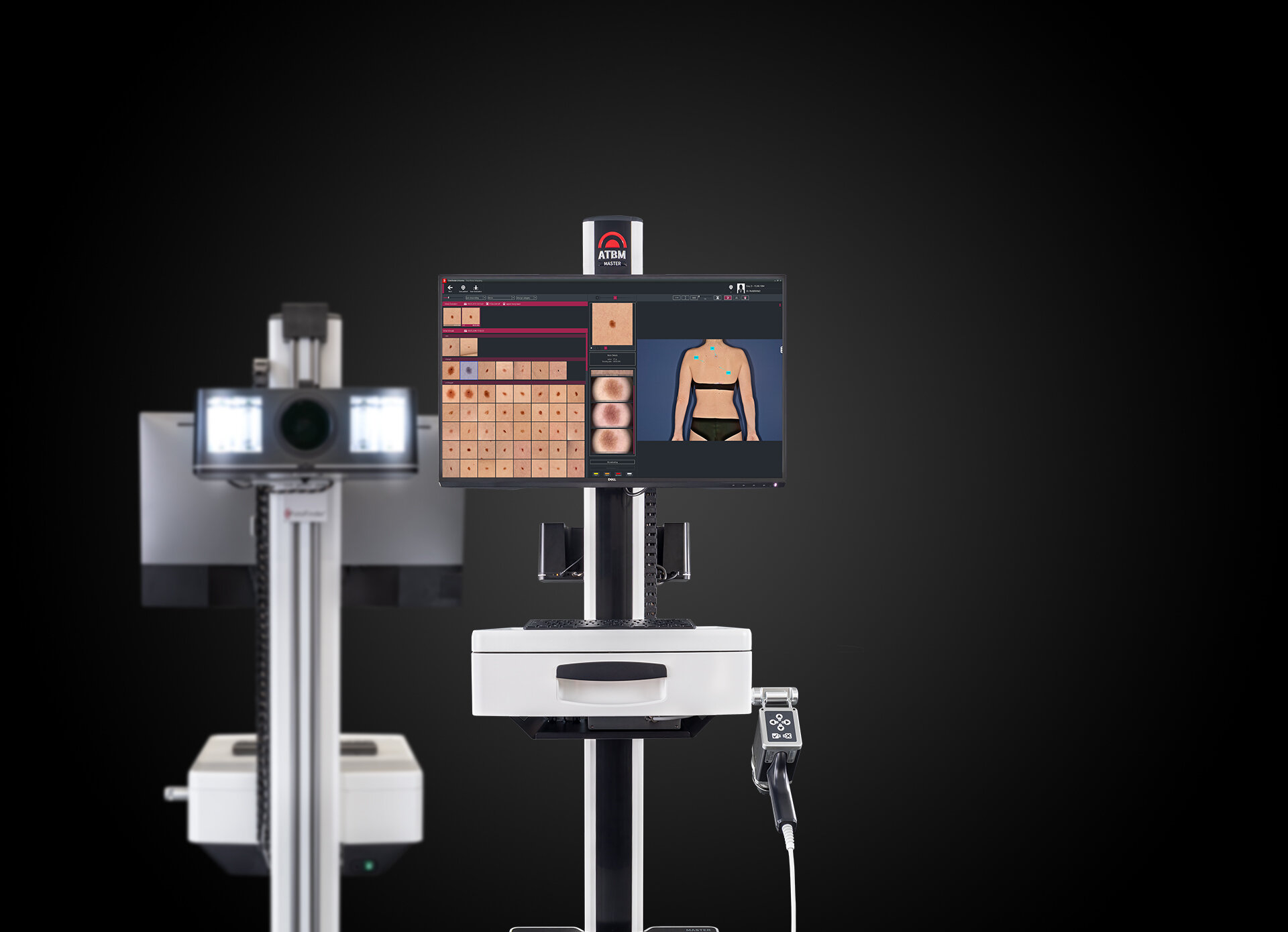
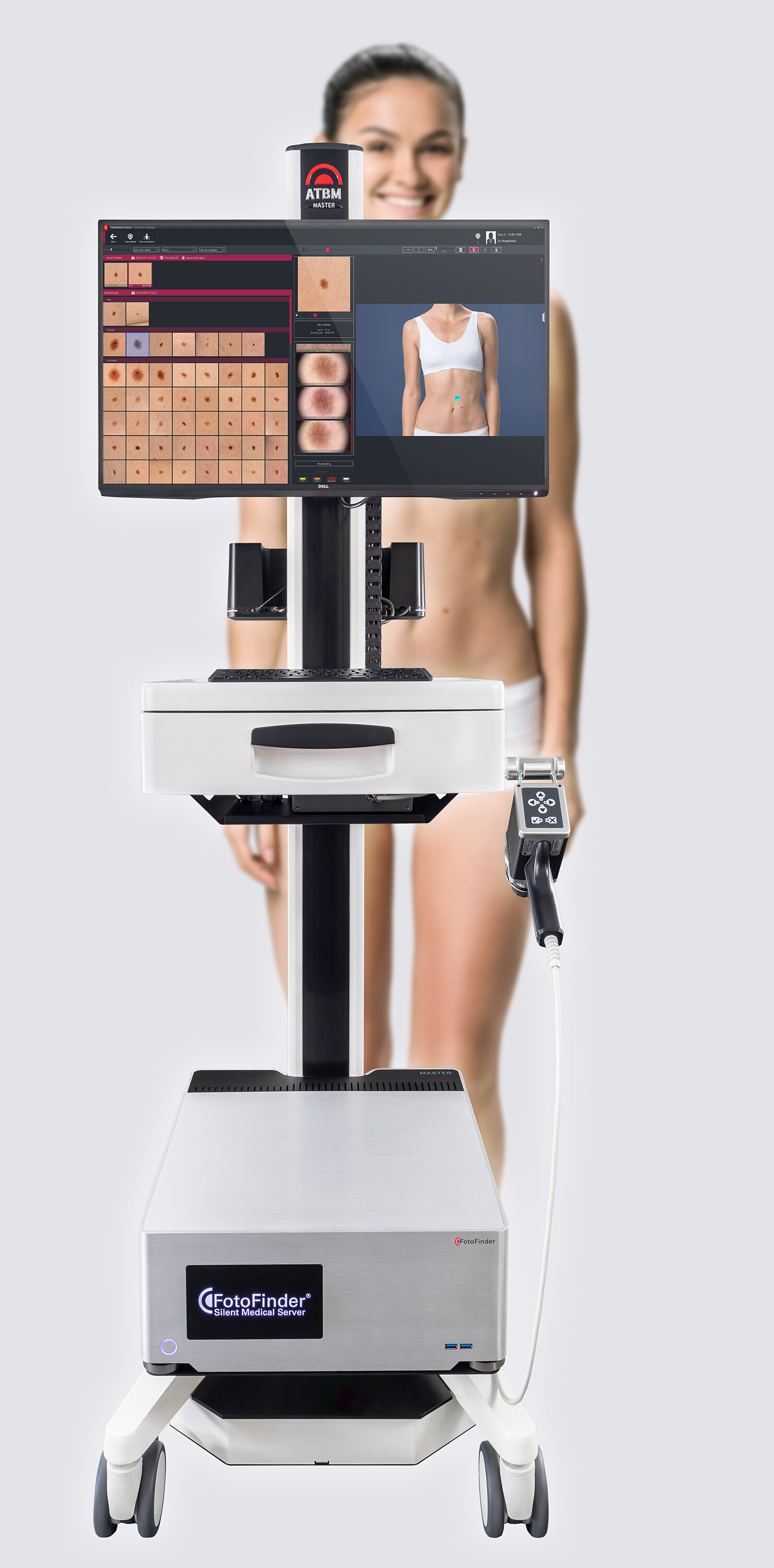
North Queensland Skin Centre utilises the Fotofinder Automated Total Body Mapping (ATBM)  System to aid in detecting new and changing lesions. It creates an accurate skin map of a patient in less than five minutes. The Fotofinder ATBM posesses an ultrahigh definition 50mp automatic camera with a polarized lens for the clearest and defined images. The camera is mounted on a computer controlled lift mechanism and moves to exact levels to take four photos of each side of a patient in quick sequence. These photos are stitched together to eliminate overlapping redundant areas and create an accurate skin map. This set of high-resolution photos are used as reference for follow up skin exams and allows the detection of new and changed moles. 

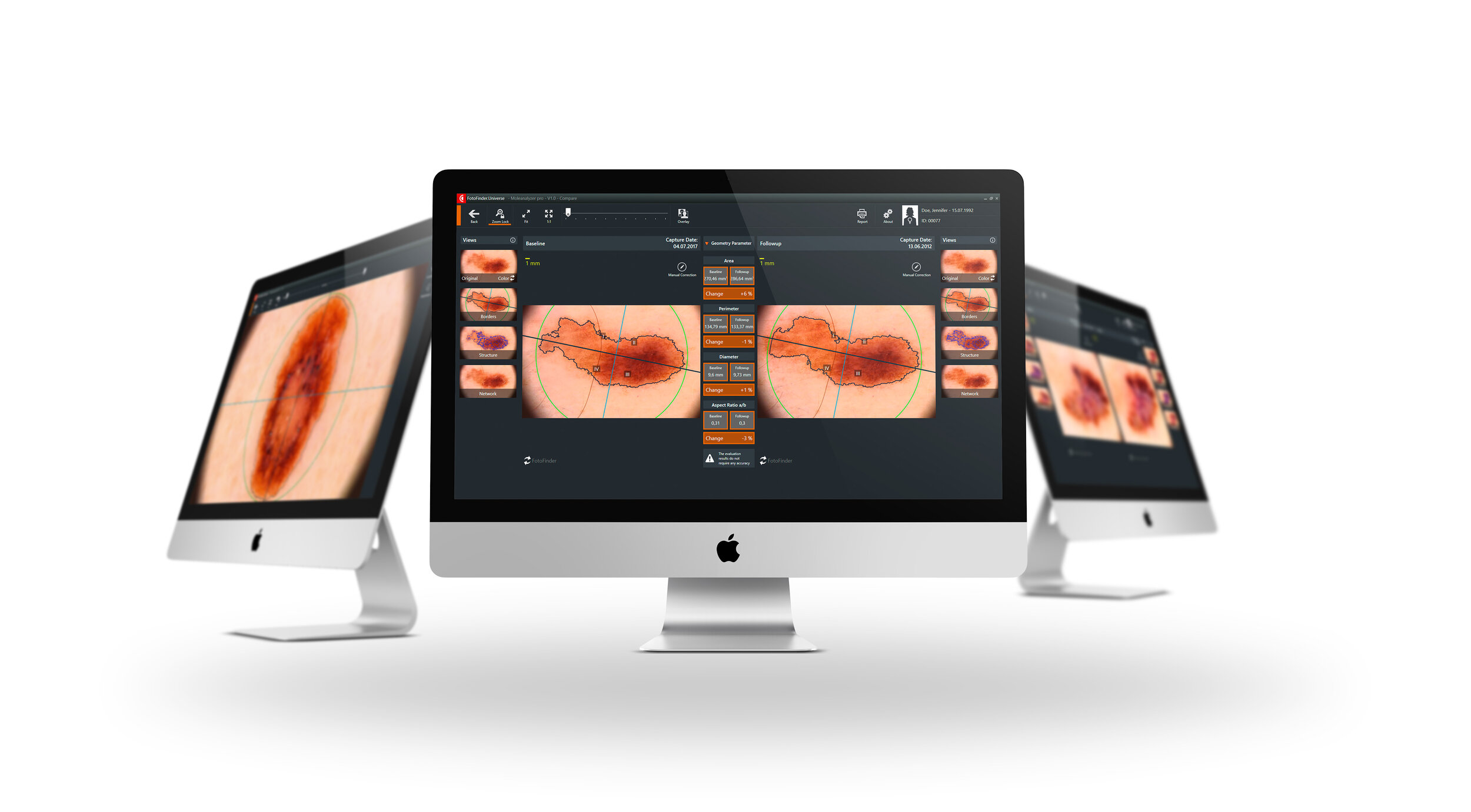
The initial scan simply acts as a baseline, the software will not detect suspicious lesions on this scan. It is on follow up scans that the benefits are really realized. By comparing baseline and follow up mole mapping photos, the Fotofinder ATBM can automatically identify new lesions and visible changes in existing moles and assist the doctor in the detection of skin cancer.

The on-screen comparison of total body photography images is significantly easier and faster then traditional mole mapping methods. This is in part due to the consistency of patient position and lighting as well as improved image definition and analytical software. This increased accuracy enabled the Fotofinder system to lay claim to identifying the smallest melanoma in the world at 0.9mm. Total body photography used to be a challenging and time consuming process. But with Fotofinder ATBM, full body photography can be done in less than five minutes

Automated total body mapping does not replace but rather is used in conjunction with a full body skin check by your doctor. By combining these two methods we are able to provide the gold standard in care for detection of melanoma and other skin cancers. Some private health insurance funds will also offer a rebate for the process.


Surgical


If your doctor is suspicious of a skin lesion but not convinced it is a skin cancer they may opt to perform a biopsy. That is to take a sample to submit to the pathologist who will look at the specimen under a microscope. This usually provides more clarity to the diagnosis and decisions can be made as to the appropriate management. There are different types of biopsies. They are performed under local anaesthetic which is injected with a very small needle.

Biopsies


pbiopsy.JPG

A Punch Biopsy uses a special tool to take a cylindrical portion of the skin lesion. A sample may be taken or if the lesion is small enough the whole lesion may be removed. Sometimes the wound is sutured. These biopsies heal very well and so the doctor may stop the bleeding and then simply dress the wound without suturing.

A Shave Biopsy takes a very thin slither of skin and leaves a wound akin to a graze or ‘gravel rash’. It is particularly useful for lesions the doctor suspects to be very thin. They heal very well with only minimal scarring and do not require sutures.

A Curette uses a tool to take a scoop of the lesion. Generally the base of the wound is cauterised with diathermy. No sutures are used and the resultant wound is a shallow crater. It heals over a week or two. It can be used to treat minor skin cancers on parts of the body where excision would be difficult for example the legs.

curette.jpg

An Excisional biopsy is used when the whole lesion needs to be removed to be submitted for examination by the pathologist. The excision is usually performed in the form of an ellipse and sutured closed. It is considered a biopsy rather than treatment as the intention is to investigate rather than treat. Some cancers like melanomas will require further treatment once the diagnosis is made.


Excision

For many skin cancers the best management is in the form of an excision. It is performed with the use of local anaesthetic which unfortunately needs to be injected with a needle and does sting a little. The doctor is careful to try to minimize the pain by using a very small needle and buffering the solution. 

excsion-1.jpg

Once the skin is numb the cancer is generally excised in the form of an ellipse or oval. It usually needs to be three times as long as it is wide to neatly bring the skin edges together and form a nice scar.There may be some bleeding but this is stopped with sutures or the use of diathermy to cauterise the vessel.

Once the bleeding has stopped the wound is closed. Often the doctor will use internal absorbable sutures. These provide a lot more strength and stop the scar from spreading. They remain inside and your body will absorb them over 3-6 months’

excsion-2.jpg

The skin edges are then brought together by more sutures at the surface. These will need to be removed in generally 7-10 days time.

With any procedure there is always a risk of infection and is usually in the vicinity of 5%. There may be bleeding but this can be managed easily. It is impossible to avoid a scar but the doctor will aim for a nice white line however scarring can be unpredictable. The specimen will be sent to be examined by the pathologist. We usually aim to remove it first time however this can not be guaranteed and occasionally it will need to be re-excised.


Flaps & Grafts

Occasionally the defect created by the removal of a skin cancer can be too large to close by suturing the skin edges together. It may require a more complex closure like a skin graft or flap.

flap 1.JPG

In the first instance a Skin Flap is used. This method borrows some adjacent skin to fill the defect. The skin is partially lifted but left attached so as to preserve the blood supply. It is then moved to fill the defect and will result in a subsequent defect. This may be closed with sutures or require a further flap to close. As it has its own blood supply and utilises neighboring skin with similar features it heals better and produces a better scar.

flap2.JPG

If a flap cannot be used then the wound can be closed with a Skin Graft. This involves removing a piece of skin from another part of the body and securing it over the defect. The graft may be split thickness which will leave a graze at the donor site or full thickness which leaves a wound that will need to be closed. Grafts have an increased risk of not taking and can have scars that are more noticeable. Often a dressing will be sutured over the top of a graft and will remain in place and need to be kept dry until taken down a week later.


Non-surgical

FLUOROURACIL & CALCIPOTRIOL CREAM


daylight photodynamic therapy

‘Sunspots’ or solar keratoses are pink scaly spots that appear on sun damaged skin. They can be irritating and appear unsightly but more concerning is that they can be a precursor to a skin cancer called squamous cell carcinoma (SCC). When there is widespread damage of solar keratoses over a piece of skin it is called ‘field change’. There are a variety of treatments available to treat field change ranging from creams to light therapies.

Currently the best evidence for clearance of solar keratosis is a combination cream of fluorouracil and calcipotriol. The two agents have differing modes of action with the fluorouracil causing an inflammatory reaction and the calcipotriol creating an immune response, that is the body learns to fight the sun damaged cells itself. The dual action means an increased clearance rate compared to other treatments. The immune response is maintained after treatment meaning there is a longer lasting effect post treatment.

The cream is applied twice a day for 4 days to the face, scalp, neck and ears and for 6 days to the forearms and backs of hands. This is much shorter than other treatments. The active substances only work on sun damaged skin so normal skin is spared. So it should be spread all over the body parts being treated, not just to the visible sun spots. By day 3 on the face and 6 on the forearms the reaction starts to become noticeable with redness and inflammation. Over the next few days it will become worse with scale, itch, heat and sometimes swelling. It will peak at day 7 or 8 on the face and 10 -12 on the arms. It will then quickly improve generally by day 10 to 14 it will have cleared.

The result is clearer skin that looks and feels better but more importantly a reduced risk of squamous cell carcinoma. It is quite a variable reaction  with some people only noticing some mild redness, they still however get an effective result. Others unfortunately can get a much bigger response with swelling, burning and pain. This is rare and though it may take a little longer to improve it will still clear. The cream will make you quite sun sensitive and potentially make the reaction worse, so it is important to protect yourself from the sun. Sunscreen can be applied over the top about 15-20 minutes later.

I n order to help with recovery it can be useful to apply a sting free ointment to keep it moist. We recommend QV Dermcare Sting free ointment. This will both soothe the skin and aid recovery. Cool compresses and face washers can also help. If there is a very large reaction that is burning and painful and not tolerated ensure you see your doctor as there are creams that can be prescribed that will help.

The cream is not available commercially. Your doctor will issue a prescription which will need to be filled at a compounding pharmacy. There are quite robust studies providing great evidence for the use of this cream. However as it isn’t available commercially it has not been taken to the TGA for approval in Australia so technically it is off label. For more information about how it works or the studies performed talk to your doctor at your next skin check or click the button below to see our Blog Piece.

DAY 7                                                                                            Day 14

DAY 7 Day 14


Daylight photodynamic therapy is a method for treating sun damaged skin to fix the sun damage to both prevent skin cancers from arising but also to regenerate the skin. Either of two creams can be used, Methyl aminolevulinate (Metvix) or 5-Aminolevulinic acid (5ALA). These creams are photosensitive so activated by light. They are preferentially taken up by the sun damaged cells, so spare normal skin. Visible light then turns them into toxic metabolites which causes the damaged cells to die off and the healthy cells regenerate.

Sunscreen is initially applied then the cream to affected areas. Sunscreen will block the damaging UV radiation but not the visible light from the sun. The patient then goes outside into direct sunlight for 2 hours, then goes inside to wash off the cream and stay out of the sun for the rest of the day. By that evening there is usually a mild reaction of redness and occasionally crusting, it is not painful. It is important to moisturise regularly. By day 3 the reaction has peaked and it is normally back to normal after 7 days.

If the sun damage is particularly bad with crust and scale there may be a need to have a pretreatment with a salicylic acid ointment to remove the scale. It can be performed when overcast but not if it is dark and raining.

Day 3                                                                                            Day 7

Day 3 Day 7


Cryotherapy

Cryotherapy can be used to treat precancerous lesions called solar keratosis, some superficial skin cancers as well as remove benign lesions like warts. Liquid nitrogen which is very cold at -273OC is sprayed onto the lesion. This causes frostbite and freezes the cells. When they thaw the cells burst and die. It does cause some slight discomfort but this resolves rather quickly. It results in a burn that turns into a scab and heals in a week or two. Occasionally it will leave a white mark in its place.

As the liquid nitrogen only penetrates so far, it is restricted to superficial lesions. There is also no ability to send the specimen for analysis by a pathologist, so the treating doctor needs to be sure of what they are treating. They may choose to perform a biopsy beforehand.

Cryotherapy_Combined.jpg

Aldara

Imiquimod is a cream that is also better known by its brand name of Aldara. It was initially developed to treat genital warts but since has gained approval for the treatment of sun damage called solar or actinic keratosis as well as certain types of skin cancer called basal cell carcinoma. It is available via a prescription from your doctor.

It works by stimulating the immune system to attack the damaged or tumorous cells and this will cause a localised reaction and inflammation. This normally consists of redness, swelling, blistering, crusting and sometimes pain. As the it works by stimulating the immune system occasionally patients will experience ‘flu like symptoms’ including muscle aches, lethargy and headaches.

Before treating basal cell carcinoma the diagnosis is normally confirmed first via a biopsy. If it is appropriate then the usual regime is once daily for 5 days a week for 6 weeks applied to the lesion and small amount of surrounding skin.  This treatment is usually able to be subsidised via the PBS.

Imiquimod can also be used for field change of sun damage. This is not subsidized by the PBS, a script will normally cost around $65. The cream is applied the area of sun damage once a day 3 days a week for a total of 4 weeks.

There is currently research being performed into the use of Imiquimod on other skin cancers including a certain type of melanoma called Lentigo Maligna.


Efudix

Fluorouracil (Efudix) cream is approved in Australia for the treatment of solar skin damage or solar keratosis as well as intraepidermal carcinoma. Solar keratosis is a precancerous lesion that can develop into skin cancer while intraepidermal carcinoma is a very early and superficial form of skin cancer called squamous cell carcinoma. Efudix causes an inflammatory reaction that kills the damaged cells but preserves normal skin. It is only available via prescription and costs about $50. 

It is applied once to twice a day for 2-4 weeks depending on the area being treated. It does result in quite a marked reaction with inflammation, redness, skin sensitivity and sometimes burning. As the treatment progresses there may be ulceration and crusting as the dead cells are shed. This is a necessary part of the treatment and evidence that it is working. After the treatment is finished skin will return to normal in about 2 weeks. It is very effective at treating sun damaged skin and returning the skin to a relatively normal state. It is important to stay out of the sun during treatment and cannot be used in pregnancy. Occasionally it can cause headaches, nausea and mouth ulcers. Your doctor will usually want to reassess you once the inflammation has settled about 4 weeks later to determine the effectiveness of the treatment and if there are any remaining lesions that will need to be treated.


Diclofenac gel (Solaraze) is used for the treatment of precancerous lesions called solar keratosis or sunspots. It is part of the non-steroidal anti-inflammatory drugs class (NSAIDS). As well as having an anti-inflammatory effect it has an anticancer effect also although how exactly is not known. It may cause a slight reaction of redness and itch but is generally well tolerated especially when compared to some other treatments. Its main drawback is that it needs to be used twice daily for 60-90 days in order to be effective.

Solaraze